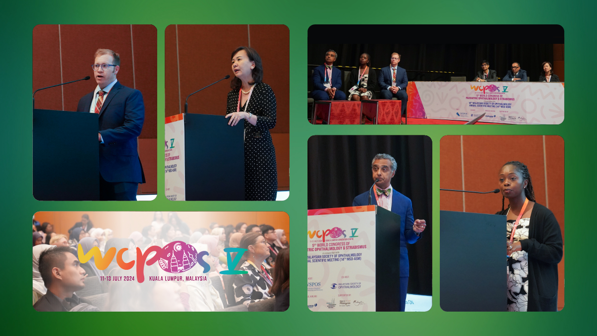
Demyelinating and neuromuscular diseases can significantly affect children’s vision and eye function. Early recognition and treatment are essential for managing these issues. At the recently held 5th World Congress of Pediatric Ophthalmology & Strabismus (WCPOS V 2024 ), experts discussed the visual challenges of these disorders and shared insights on treatment strategies to preserve and restore sight in children.
Optic neuritis with encephalomyelitis
Optic neuritis (ON), which is the inflammation of the optic nerve, can be a crucial indicator of underlying demyelinating conditions. According to the Pediatric Optic Neuritis Group, 50% of optic neuritis cases are either post-infectious or post-immunization, while the other 50% are associated with underlying neuroinflammatory disorders. Symptoms commonly linked with ON include numbness, ataxia, limb weakness and constitutional symptoms such as prolonged vomiting, hiccups or new-onset seizures, as shared by Dr. Ng Sui Yin (Malaysia).
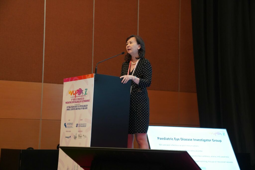
Zooming in to ON with encephalitis, she said, “There are five categories associated with ON with encephalitis: acute disseminated encephalomyelitis (ADEM), clinically isolated syndrome (CIS), multiple sclerosis (MS), pediatric neuromyelitis optica spectrum disorder (pNMOSD) with AQP4-IgG positivity, and myelin oligodendrocyte glycoprotein antibody disease (MOGAD).”
She continued to share about the clinical presentation of ADEM, which includes encephalopathy and polyfocal deficit. Usually, children with ADEM had a history of viral infection or a recent immunization days or weeks before presentation. Meanwhile, CIS is used to define white matter disease suggestive of inflammatory demyelination with symptoms lasting more than 24 hours but that do not meet the criteria for a diagnosis of ADEM, neuromyelitis optica or multiple sclerosis, and cannot be explained by other ethology. As for MS, the presence of oligoclonal bands in the spinal fluid is a good marker. On the other hand, pNMOSD and MOGAD are quite rare.
She concluded by saying that the diagnostic workup for children with ON includes MRI of the brain and orbits with contrast, MRI of the spine with contrast (especially for patients with recurrent disease or other neurological symptoms), serology testing for neuromyelitis optica and myelin oligodendrocyte glycoprotein (MOG) antibodies, and cerebrospinal fluid (CSF) analysis for patients with a high index of suspicion for multiple sclerosis.
Treatment guidelines for pediatric demyelinating disorders
Pediatric demyelinating disorders are a group of conditions affecting children in which the protective covering of nerve fibers, called myelin, is damaged, impairing nerve function. Multiple sclerosis (MS), a common pediatric demyelinating disorder that involves formation of plaques in the central nervous system, is generally milder than adult cases and occurred in 3-5% of all MS cases, noted Dr. Prem S. Subramanian (USA).
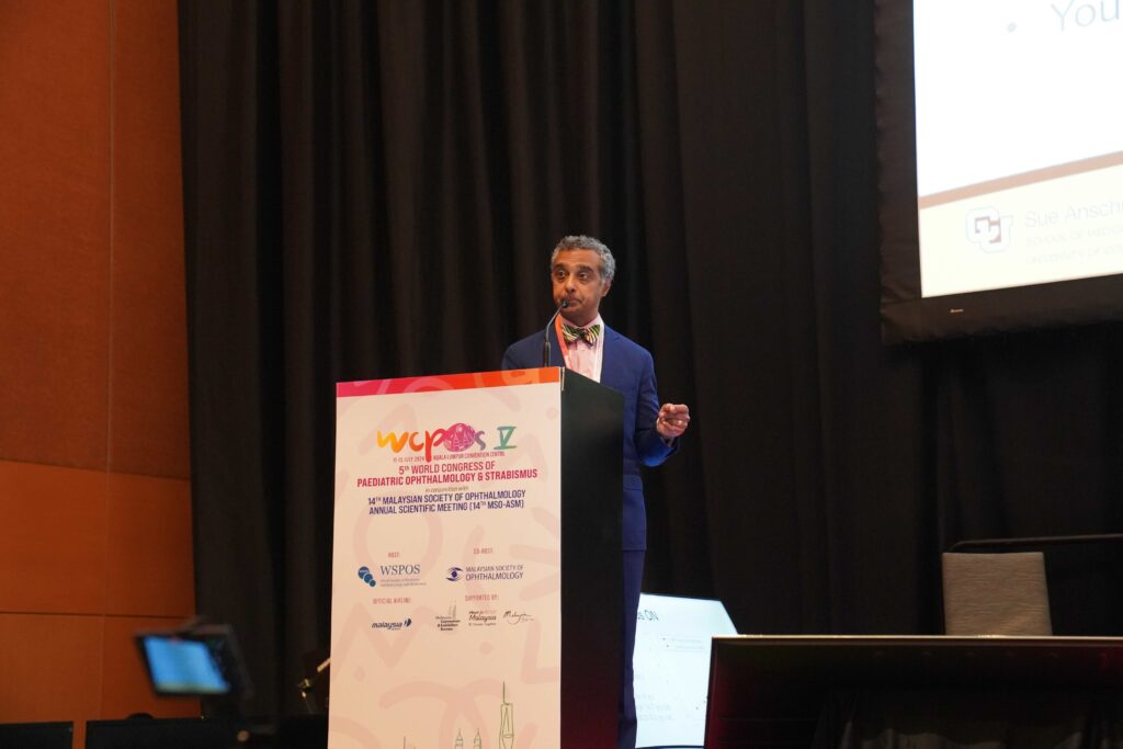
He mentioned that the acute treatment of pediatric MS often involves the use of intravenous corticosteroids given at 20-30 mg/kg/day for 3-5 days. “Patients are typically given oral taper especially if they have incomplete resolution of symptoms. Plasma exchange (PLEX) is only used in refractory cases that are not improving, and steroids are repeated for relapse,” he said.
Meanwhile, chronic treatment varies by disease. “Fingolimod is the only FDA-approved medication for children with MS and is considered superior to weekly interferon-beta,” he said.
In pediatric MOG-antibody-associated diseases (MOGAD), acute treatments include intravenous methylprednisolone, PLEX, and intravenous immunoglobulin (IVIG); while chronic treatment involves long-term oral steroids, rituximab, mycophenolate mofetil and IVIG. The goal is to promote recovery of function and prevent relapse, he emphasized.
Pediatric cranial nerve palsies
Cranial nerve palsies encompass a spectrum of conditions, including supra-nuclear control disorders, nuclear lesions, nerve palsies, orbital lesions and myopathies.
Dr. Virender Sachdeva (India) highlighted that the definitive treatment is surgery, and it is advisable to observe for recovery over a period of six months to one year. Prognosis can range from fair to poor. Better outcomes are observed in cases of congenital palsy and inflammatory causes, while recovery from trauma or compressive causes is usually poor, he noted.
“In pediatric cases, diagnosis is more difficult because detailed history may not be available and medical examination may not be possible. Children may not be cooperative. We may need to rely on saccadic velocities and magnetic resonance imaging (MRI) may be mandated,” he said, adding that neuroimaging will be indicated if there is incidence of trauma, associated headaches and neurological signs, associated disc edema, associated acquired nystagmus, worsening of observation or no signs of spontaneous recovery.
In conclusion, Dr. Sachdeva highlighted that cranial nerve palsies are not uncommon. “They can be congenital or acquired. We should try to localize and look for accompanying signs to help us pick up myopathy. We should rule out primary myopathies. Neuroimaging and ancillary testing are important. Early interventions can help to prevent amblyopia,” he stressed.
Pediatric neuromuscular junction disorders
Myasthenia gravis (MG) is an autoimmune disorder affecting the neuromuscular junction (NMJ). It is the most common NMJ disorder in children and most often presents with symptoms such as variable ptosis and strabismus, noted Dr. Ellen Mitchell (USA).
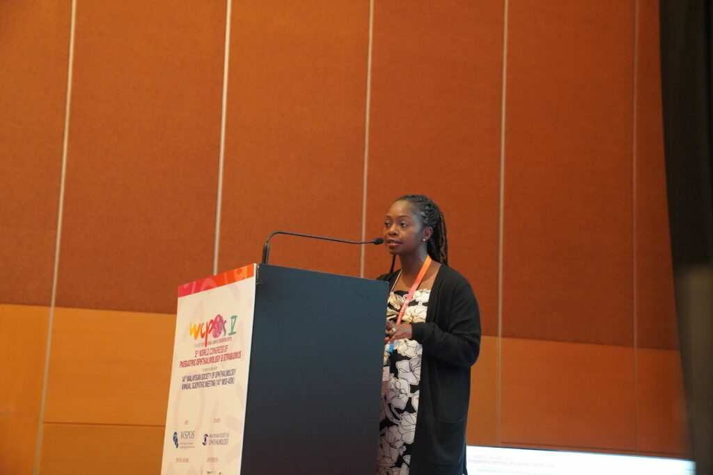
“Pediatric myasthenia gravis comes in three forms: congenital myasthenic syndromes, transient neonatal myasthenia, and juvenile myasthenia gravis,” Dr. Mitchell reminded the audience.
She emphasized that congenital myasthenic syndromes can present in infancy, and a genetic diagnosis is necessary to tailor treatment. On the other hand, transient neonatal myasthenia gravis occurs in 5-30% of infants born to mothers with MG and presents after birth with symptoms such as a weak cry, poor sucking, respiratory distress and generalized hypotonia. Meanwhile, juvenile myasthenia gravis is characterized by fluctuating fatigue of skeletal muscles in patients aged 0-18 years. Symptoms include fatigable ptosis, variable strabismus, and general weakness.
“These infants may experience flaccid paralysis and require close monitoring for respiratory compromise. They typically respond well to treatments like neostigmine and plasma exchange. Once autoantibodies are cleared, symptoms usually resolve,” she explained.
Furthermore, Dr. Mitchell also discussed Lambert-Eaton myasthenic syndrome (LEMS). “The diagnosis of LEMS should be considered in children with unexplained progressive proximal muscle weakness and a negative evaluation for more common causes. Some clinical symptoms of LEMS, such as ptosis, overlap with those of other myasthenic syndromes, most commonly MG, which can lead to misdiagnosis or delayed diagnosis,’ she emphasized.
Pediatric ocular myasthenia
Zooming in on ocular myasthenia gravis (OMG), Dr. Venkateshwar Rao (India) noted that OMG occurs in 10-35% of all childhood MG cases and has a higher prevalence in Asians. The most common manifestation is ptosis (90%). Diplopia and strabismus occur in 45% of cases, complete ophthalmoparesis in 24% of cases and 40-50% of cases convert to generalized myasthenia gravis, he noted.
Dr. Rao shared that clinical signs of OMG include fatigability on sustained upgaze, and Cogan lid twitch. The ice test (80% sensitivity), rest test, edrophonium (Tensilon) test and neostigmine test, and laboratory testing are useful in diagnosing the condition.
In terms of treatment for OMG, anticholinesterase drugs are the mainstay. Most patients need immunosuppressive therapy as well. Early immunosuppression helps to prevent progression.
“All patients with MG and thymoma should undergo thymectomy. Strabismus surgery can be performed for stable deviation, and full spontaneous remission does occur in the majority of these patients,” said Dr. Rao.
Editor’s Note: Reporting for this article occurred at the 5th World Congress of Paediatric Ophthalmology & Strabismus (WCPOS V 2024) from 11-13 July in Kuala Lumpur, Malaysia.