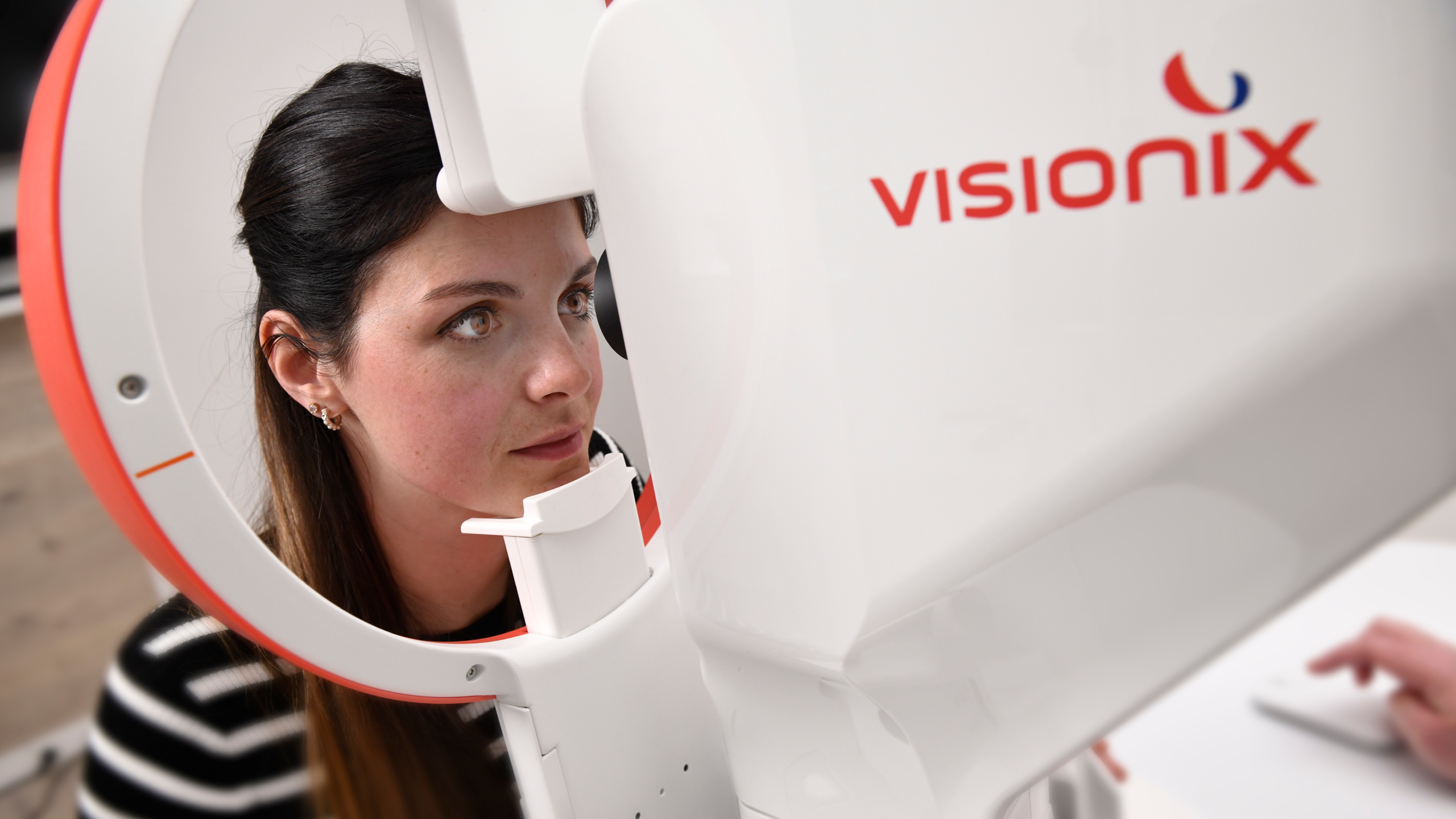
Sponsored by Visionix
Why settle for a diagnostic snapshot when you could have the full gallery? The Optovue Solix OCT/OCT-A offers high-def imaging, fast scans and smart analytics, giving you more than just a pretty picture. It’s time to see the whole story.
In photography, there’s a clear line between a quick smartphone snap and an image captured with a professional camera: the depth, the crisp detail, the ability to capture what most eyes miss. The same goes for eye care diagnostics. The Optovue Solix OCT/OCT-A (Visionix; Normandy, France) delivers that same high-definition clarity, bringing professional-grade imaging straight into your practice.
The device marks a meaningful step forward in spectral-domain OCT (SD-OCT) and OCT angiography (OCT-A). High-resolution images? Check. Lightning-fast scans? Check. A broader field of view? Also check. This isn’t a dramatic reinvention, but it is a well-calibrated refinement that gives you more information with less effort.
“With the Optovue Solix, I moved from a lower density radial ring pattern to a very high density Q pattern,” said Dr. Robert Rothstein, founder of Eyenamics NY (USA).
“In addition, there’s deep learning segmentation and improved artifact removal. This gave me better repeatability, reproducibility and more trustworthy trend data,” he noted. “I make decisions with greater confidence and fewer rescans…it’s almost three times the improvement.”
The Optovue Solix’s 120,000 A-scans per second speed helps deliver detailed images of the retina, choroid and anterior segment, without the motion artifacts that used to haunt OCT scans. The result? Fewer rescans, better efficiency and clearer images you can count on.
Seeing more, seeing smarter
The system’s 12 mm by 12 mm widefield en face OCT gives you a panoramic view of the retinal and choroidal vasculature. With it, you’re no longer boxed into the central macula. Now, you can assess the periphery with the same clarity a wide-angle lens brings to a landscape shot.
“Recently, I saw a patient who had pseudotumor cerebri, but I also suspected posterior choroiditis. You could see on the OCT and fundus photos evidence of multifocal choroiditis,” Dr. Rothstein recalled.
“The Optovue Solix can actually look at abnormalities of the retina found on fundus photography, especially on wide field fundus photography, and then see a cross section of it on the OCT. The optic nerve scans are now 6 mm by 6 mm, so you have a higher density and less interpolation.”
The Optovue Solix’s AngioVue® technology gives you a dye-free, non-invasive look at microvasculature, while AngioAnalytics™ translates those visuals into hard numbers (like vessel density, flow area, and the foveal avascular zone). The combination delivers both visual and quantitative insights to support more informed decision-making.
And this visual versatility extends beyond the retina. The Optovue Solix handles anterior segment imaging with the same precision. Corneal epithelial, stromal and pachymetry maps help inform keratoconus management and refractive surgery planning. Its anterior chamber angle assessment adds yet another layer, visualizing angle structures and measuring them with the clarity of a macro lens capturing every fine edge.
Speed without sacrifice
In today’s busy practice environment, every second counts. With rapid scanning speeds and smart automation, the Optovue Solix helps eye care pros stay efficient without cutting corners on image quality.
Thanks to its built-in Motion Correction Technology (MCT), the system automatically smooths out patient movement (yes, even the fidgety ones), minimizing the need for frustrating rescans. And with FastTrac™ eye-tracking, it keeps the image crisp by adjusting for eye motion mid-scan.
“The Optovue Solix is faster and requires fewer repeats. It certainly cuts back chair time, so we could see more patients,” said Dr. Rothstein. “The Optovue Solix now has 120,000 A-scans versus Avanti, which was 60,000. In terms of workflow, it’s easier to see patients faster, and it’s certainly more reliable testing.”
Think of it like a high-end camera with image stabilization and autofocus. You’ll capture what you need, fast and sharp, right from the first click.
A big-picture view of glaucoma
Like a seasoned photographer balancing lighting, composition and timing, the Optovue Solix takes a similarly thoughtful approach to glaucoma management, offering a big-picture view with sharp attention to detail. By combining structural and vascular measurements, it helps clinicians see more of the disease process…and see it earlier.
“With the Optovue Solix, you can now really correlate RNFL and ganglion cell loss with vessel density loss. We now have another indicator, another biomarker, so to speak, to follow when monitoring glaucoma patients,” said Dr. Rothstein.
“The Optovue Solix is actually much more sensitive in picking up changes that might not have been picked up on the Avanti. You can pick up changes in the RNFL and the ganglion cell complex, and you might be able to pick up earlier pathology in a patient with suspected optic neuropathy, in particular. Or in glaucoma patients, you might pick up earlier progression.”
And then there’s the Hood Report. This feature overlays OCT data with visual field results, making it easier to connect the dots between what you see and what the patient experiences. Think of it as the diagnostic version of a photo essay that reveals not just the headline image, but the full narrative.
Developing the big picture
Investing in a system like the Optovue Solix isn’t just about upgrading your tech, it’s about leveling up your entire practice. While the clinical perks are clear, the financial benefits are worth a closer look, too.
With its all-in-one imaging capabilities, the Optovue Solix can help you consolidate testing, streamline workflows and potentially boost reimbursement. It’s a win-win: fewer tests for patients, more efficient throughput for you. And when you’re spotting disease earlier and managing it more effectively? That translates to better outcomes, happier patients and maybe even a few glowing reviews.
“Where the OCT can become a bottleneck, when it’s easier to see these patients and scan them with more reliability, we can increase the volume. Patients can be satisfied because there’s less chair time,” said Dr. Rothstein. “Regarding reimbursements, the OCT-A is now being reimbursed at a higher fee [in the United States], and that’s certainly a gain for the practice.”
Think of it like this: a professional photographer doesn’t settle for a smartphone snap. They invest in gear that captures every nuance. The Optovue Solix is that high-performance camera for your clinic, delivering crystal-clear, wide-angle views paired with powerful analysis tools that help you catch what others might miss.
In a field where details matter, the Optovue Solix OCT/OCT-A doesn’t just help you diagnose, it helps you tell the whole story of ocular health, one expertly captured scan at a time.
Meet the experts
Curious about the newest Optovue Solix innovations in action? Want tips straight from the surgeons who use the Optovue Solix day in, day out? Swing by the Visionix booth (Stand B4.116) at ESCRS on September 13 and 14, 2025. Join the experts for lively, hands-on booth talks packed with insights, real-world cases, and clever ways to elevate your diagnostics. Discover how bringing true anterior OCT to Visionix’s renowned posterior technologies can help you see everything, crystal clear.
Editor’s Note: This content is intended exclusively for healthcare professionals. It is not intended for the general public. Products or therapies discussed may not be registered or approved in all jurisdictions, including Singapore.

