From dislocations to inverted ICLs, this session revealed hard-won lessons and clever saves in phakic lens surgery.
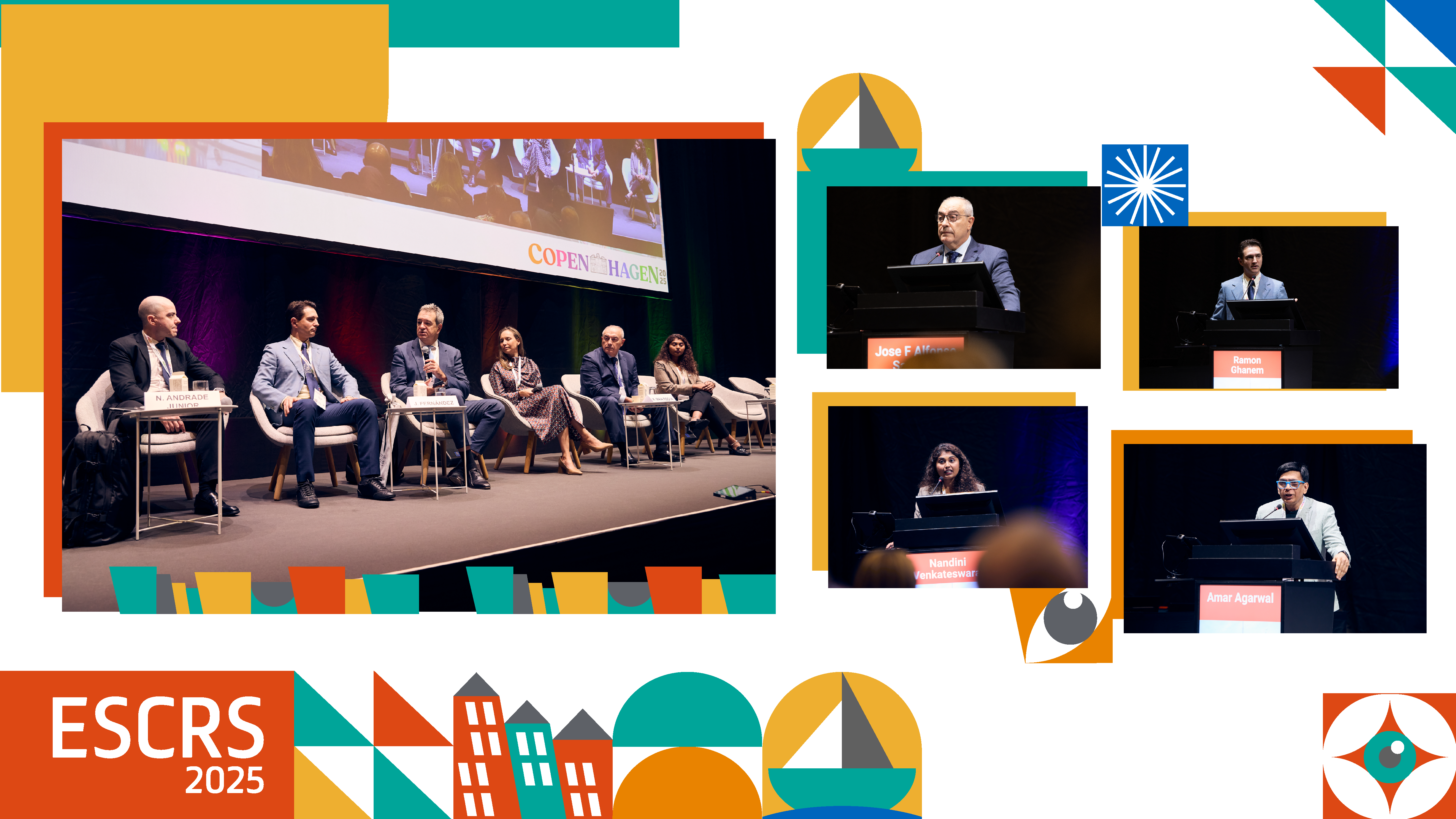
Under the bright lights of the Bella Center in Copenhagen, a Day 1 session of The Global Refractive Summit on managing complications in phakic lens surgery set an energetic tone for the 43rd Congress of the European Society of Cataract and Refractive Surgeons (ESCRS 2025).
Chaired by Drs. Newton Andrade Junior (Brazil), Joaquín Fernández (Spain) and Karolinne Maia Rocha (United States), the session wove together three decades of experience, evolving implantable collamer lens (ICL) technology and inspiring “war stories” from a truly global faculty of phakic lens experts.


Anterior chamber pearls (and pitfalls)
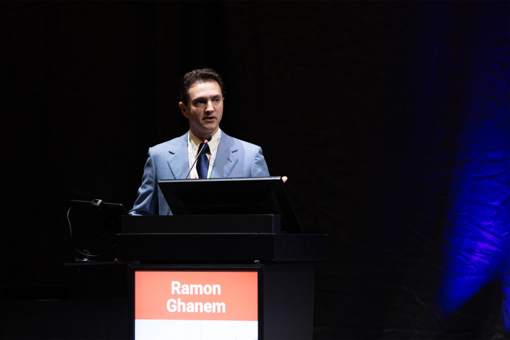
Opening the clinical arc, Dr. Ramon Ghanem (Brazil) traced a 30-year perspective on Ophtec’s (Groningen, The Netherlands) iris-fixated Artisan/Artiflex aphakic IOLs. His talk was practical and historical in equal measure.
READ MORE: Ophtec’s Phakic IOL for Presbyopia Receives CE Mark Approval
On enclavation, he stressed precision: “It’s important to note that you need a proper amount of tissue enclavated. If you have a poor enclavation, you may lead to iris atrophy and late IOL dislocation.” For re-fixation, he favors two paracenteses and Ophtec’s VacuFix system to aspirate stroma and “increase the tissue inclavated” if stability is doubtful.
Endothelial safety was his through-line. He highlighted the current ≥3.0 mm aqueous distance rule, reminding colleagues that with proper technique, outcomes remain steady—“only 12 percent median endothelial cell loss” at ten years with Artiflex. Still, he cautioned against complacency: hyperopic cases, rubbing or shallow chambers can tip the balance.
How about cataract risk? Rare but real. Patients over 40 and extreme axial length remain red flags. His surgical choreography for combined care was crisp: “Our usual technique is a 2-site explantation and 2-site phaco. But don’t try to do this in the same incision. You have an unstable chamber and risks are increased.”
And a closing caution every anterior-segment surgeon knows to their core: “Avoid small iridotomies.” One patient’s pupillary block became the worst case in his clinic; the fix (and the lesson) were indelible.
The posterior chamber perspective
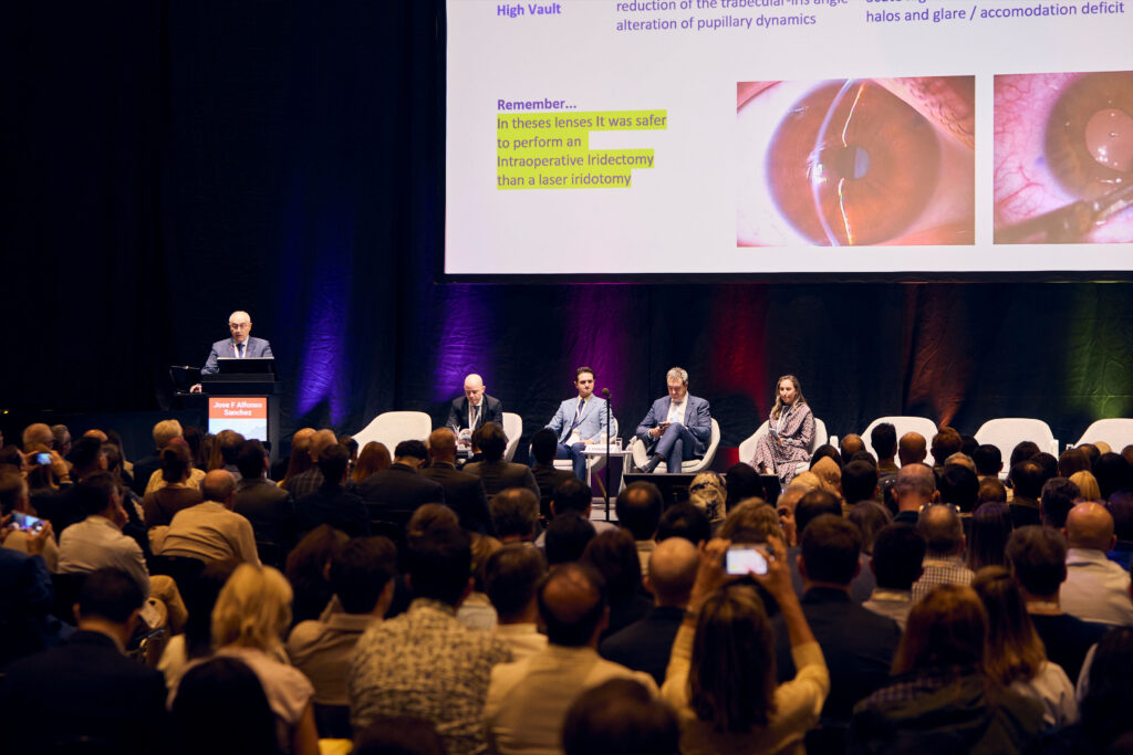
Dr. José F. Alfonso Sánchez (Spain) brought clarity to the slippery concept of vaults in posterior chamber IOLs. “Are we talking about central or peripheral vault, nasal or temporal, static or dynamic, initial or final?” he asked, underscoring the nuance.
Sizing mistakes cause problems. “In general, a low vault is due to lenses that are too small and a high vault to lenses that are too large.” With central-port ICLs, true pressure crises are uncommon unless the port is occluded, but high vaults can still alter pupil dynamics and induce halos.
WATCH NOW: Phakic IOL Experts on Implantation Candidacy and Power Calculations
To tame the outliers, he advocated intraoperative anterior-segment OCT: “If the intraoperative central ball is greater than 600 microns, we recommend rotating the lens towards the vertical meridian. If the value is less than 200 microns, we don’t move it.”
His long-term reassurance was striking: “With the anterior chamber IOLs with a central port, a hole less than 250 microns doesn’t produce cataracts in 15 years of follow-up.” On IOP, steroids still explain most early spikes, while pigment dispersion syndrome sits at the intersection of iris configuration and biomechanics.
In predisposed patients, he argued, the ICL can actually normalize iris contour and reduce IOP over time.
Keratoconus: Who, when and how
Dr. Nandini Venkateswaran (United States) reframed the room’s mindset around ICL implantation in keratoconus: start by picking winners. “Choosing appropriate candidates is going to set you up for success,” she said, and the non-negotiable is stability—“you need to ensure the underlying keratoconus and the refractive error are stable.”
Serial tomography and refractions are her compass; any whisper of progression lowers her threshold for cross-linking before refractive surgery.
Expectation-setting matters as much as measurements. She counsels that postoperative acuity aims to mirror best-corrected spectacles, not scleral-lens performance; trial-framing in clinic helps patients “feel” the goal.

She also doesn’t shy from the messy middle: aberrated optics, glare/halo risk, the possibility of refractive surprise and the reality that some will still use readers—or even glasses—after surgery.
Practical pearls included wound construction (3.0 mm, avoid going too posteriorly) and planning for floppy irides. And one piece of advice she repeats to every patient: “I always counsel them to of course not rub their eyes.” That habit alone, she warned, can accelerate ectasia, cataract or lens rotation.
WATCH NOW: CTAK by CorneaGen: A New Era in Keratoconus Care
Evolution of ICL technologies
Dr. Ricardo Nosé (Brazil) looked at the rapid evolution of ICL design. He reminded surgeons that reversibility is still the phakic IOL’s greatest strength. The current EVO-style designs sharply reduce cataract risk seen with older non-port models, but he echoed a common chorus: “The most important challenge is patient selection.”
Sizing accuracy is the fulcrum—white-to-white, anterior chamber depth, specular microscopy, ultrasound biomicroscopy and contemporary AI-assisted calculators, all help avoid vault extremes.
When complications do occur—say, a postoperative pupillary block or an unhappy vault—these lenses give surgeons options: “The good thing is we can also remove the IOL.”
Looking forward, he highlighted preloaded injectors, new biomaterials, presbyopic optics and hybrid bioptics combining ICL with corneal procedures and intrastromal rings.
READ MORE: Phakic Forward: Evolving the Space Between Laser and Lens
Same-day, both eyes? A data-driven case
Dr. Andrea Russo (Italy) addressed the “taboo” of immediate sequential bilateral ICL (ISBICL). With myopia affecting nearly 60% of the world, he asked: “Why not consider the ICL?” After all, bilateral same-day cataract, LASIK and SMILE are already mainstream in many centers.
In a multicenter experience across Italy, London and Belgium, he reported strong safety and refractive accuracy—“89% within half a diopter and 96% within 1 diopter” when keratoconic eyes were excluded.1
On vaults, Dr. Russo reframed perfectionism: “The ICL is very much forgiving.” While 250–750 µm is “optimal,” he argued real-world tolerance is wider and should be constrained by angle status more than a single vault number: He also reminded surgeons to be patient, since vaults often settle as haptics find their position.
WATCH NOW: Dr. Dong-Hoon Lee on Surgical Pearls from Running an ICL-Only Clinic
Looking ahead, he highlighted that while manufacturer nomograms are improving, machine-learning predictors that integrate lens rise, anterior-segment OCT metrics and broader biometry have increased sizing accuracy from the high 60s to over 90% in their experience.
“Take advantage of these machine learning formulas because they are way more precise than the one coming from the company.”
The good, the bad and the ugly—and the save
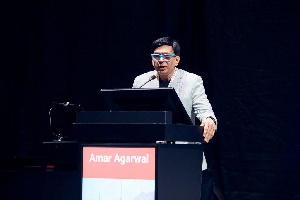
Prof. Amar Agarwal (India) framed his session with a movie reference: “I’m showing you the good, the bad and the ugly in phakic IOLs,” he said.
He began with the good: the straightforward steps of ICL implantation. “Important is the orientation. Once you have the orientation right, you grasp and pull with the duckbill forceps. Previously we used to just push it, and inverted ICL would be the result in some cases.”
But as he put it, “I really wish everything would work so nicely and so simple. But invariably it does not. So let’s move to the bad and then the ugly.”
In the bad category, Prof. Agarwal walked the audience through cataracts, inverted ICLs and stuck injectors. For inversion, his team published a paper on Glued Intrascleral Haptic Fixation of an Intraocular Lens Technique using viscoelastic and a disco cannula to flip the ICL in situ.2 Still, complications sometimes escalate. One stuck injector left him with a torn ICL.
The ugly stories were even more complex: a one-eyed patient with endothelial decompensation needing PDAC endothelial keratoplasty; an anterior chamber phakic IOL with peripheral anterior synechiae; a torn anterior capsule that dropped an IOL into the vitreous. In each case, he showed creative saves—glued IOLs, single-pass four-throw pupilloplasty, handshake techniques and even trocar-assisted iridodialysis repair.
READ MORE: Perfect Surgical Saves and Lessons from the Masters
Turning complications into manageable challenges
In the end, the session echoed a reassuring theme: most phakic IOL complications are preventable, many are reversible and nearly all are manageable with planning, precision and persistence.
From pearls on patient selection to inventive saves in the toughest cases, the playbook that emerged in Copenhagen was less about fear of complications and more about confidence in recognizing, preventing and—when needed—rescuing them.
Editor’s Note: The 43rd Congress of the European Society of Cataract and Refractive Surgeons (ESCRS 2025) is being held from 12-16 September in Copenhagen, Denmark. Reporting for this story took place during the event. This content is intended exclusively for healthcare professionals. It is not intended for the general public. Products or therapies discussed may not be registered or approved in all jurisdictions, including Singapore.
References
- Russo A, Filini O, Savini G, et al. Predictability of the vault after implantable collamer lens implantation using OCT and artificial intelligence in White patient eyes. J Cataract Refract Surg. 2023;49(7):724-731.
- Narang P, Agarwal A. Glued intrascleral haptic fixation of an intraocular lens. Indian J Ophthalmol. 2017;65(12):1370-1380.