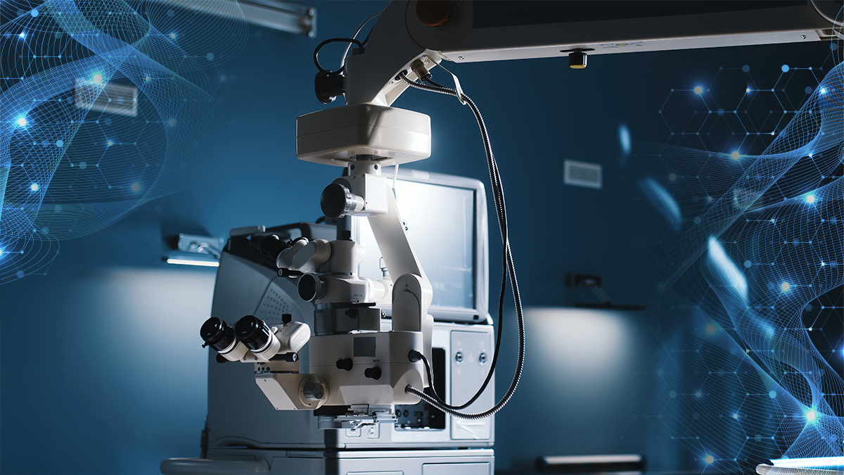
A panel of experts in Los Angeles argued that the time is now for the digital OR transition—for your patients, your clinic and your health.
If a panel of the United States’ leading anterior segment surgeons are to be believed, there’s never been a better time to go digital in the operating room.
In a Day 2 symposium at the American Society of Cataract and Refractive Surgery Annual Meeting (ASCRS 2025), Drs. Paul Singh, Julie Schallhorn, Eric Rosenberg, Robert Weinstock and more made the case for digital surgical visualization systems.
The arguments went well beyond claimed benefits to surgical efficiency; improved ergonomics, enhanced visualization and expanded functionality were among the top reasons for switching as experts discussed their transition experiences and the ample, tangible benefits of digital operating rooms.
WATCH NOW: Digital OR Series with Dr. Karen Glandorf & Dr. Nobert Pesztenlehrer
The economics of ergonomics
Dr. Deepinder Dhaliwal opened the session with a sobering reminder of the occupational hazards facing ophthalmologists and their spines.
After experiencing a severe disc herniation that nearly ended her career, ergonomics became Dr. Dhaliwal’s passion project—and for good reason.
“Musculoskeletal disorders are such a problem in our profession because there’s a high risk of repetitive tasks, tasks that require fine motor skills and prolonged maintenance of awkward body positions,” she explained, noting that surveys show up to 81% of ophthalmologists report pain in their neck, back or shoulder.1
The traditional operating microscope, with its rigid positioning requirements, is a primary culprit, according to Dr. Dhaliwal. “This is why we have so many issues with our necks,” she said. “For every inch that the head moves forward in space, the relative weight of a spine increases by 10 pounds,” putting enormous strain on surgeons over long hours in the OR.
“You gotta think about ergonomics and posture every day. We all need to strengthen our core lower back. We are elite athletes in a way, right? And so we really should take care of our bodies,” she emphasized. “We need to do modifications for the rest of your life to prevent further injury.”
Dr. Paul Singh, who transitioned to digital visualization after cervical fusion surgery, demonstrated objective postural improvements.
His before-and-after photos showed dramatically improved shoulder-head alignment when using 3D heads-up displays. “If you look at my neck position, can you see how I’m tilted? I’m twisting my head to the right. But with the correct posture, I turn and now I’m actually more straight and square with the monitor.”
For Dr. Singh, the importance of proper setup is critical, even when it goes against conventional wisdom. “The key is not to actually have to sit right across the oculars,” he said. “We are taught that we have to sit right across the oculars because that’s how we had to use them. Now with your freedom from yourself and the oculars, you can sit anywhere you want to.”
Protecting patients with the digital OR?
But there’s more to the digital OR than ergonomics— it’s also about what Dr. Eric Rosenberg believes are the superior visualization capabilities that come along with the switch—with less potential damage to patient eyes.
READ NOW: Real-World Stories in Navigating Obstacles to Digital Transformation
Reading from the guidelines of the International Commission on Non-Ionizing Radiation Protection, he revealed that at 50% light intensity with the typical center red reflex light, “the time to operate is 3.7 minutes of direct light intensity.”
“I guarantee you nobody up here is doing it in 1.1 minutes, and that’s how long they recommend to expose the eye to light with all three lights on,” Dr. Rosenberg warned.
His research suggests that reducing light exposure by using digital systems not only improved patient comfort, but produced measurable clinical benefits. “Decreasing the light at the time of cataract surgery actually decreased the rate of cystoid macular edema (CME) in diabetic patients 9-fold, and in regular patients, decreased the rate of CME 4-fold.”
In side-by-side comparisons, Dr. Rosenberg also demonstrated how digital systems operating at just 5% light intensity produced superior visualization. “When you look at the hydrodissection step, you might feel very unsafe performing the surgery. Whereas [with digital OR technology], the screen is lit up like a Christmas tree, and we can see exactly what we’re doing.”
With the infrared visualization available in some digital OR suites, Dr. Rosenberg challenged conventional thinking: “Why are we staying on the visible light spectrum?” he asked, showing how infrared technology shows through dense cataracts and other visualization issues that traditional technology has trouble with.
Intraoperative OCT integration
Dr. Julie Schallhorn believes that the intraoperative optical coherence tomography (OCT) integration with digital systems provides heretofore unimagined capabilities. “OCT is a great way of doing an optical section of the eye,” she explained. “It gives us a type of view that we’ve never been able to have before.”
This technology is particularly valuable for complex cases, according to Dr. Schallhorn. She demonstrated how intraoperative OCT helped her complete a rhexis that had disappeared under corneal opacity and identified a case of epithelial downgrowth that would have been difficult to visualize otherwise.
For secondary IOL implantation, Dr. Schallhorn also now relies on intraoperative OCT to ensure proper lens positioning. “I actually adjust the tilt of the lens to make it completely planar to the iris until I’m very, very happy… I do this now with every single secondary IOL.”
READ MORE: Revolutionizing the Operating Room
The educational advantage and future trajectory
Multiple presenters emphasized the teaching benefits of digital systems. Dr. Rosenberg demonstrated a platform called stytch.tv that enables worldwide live streaming of surgeries with interactive capabilities–-all made possible by the digital OR.
Dr. Singh echoed this with his discussion of the benefits for staff—especially when they can see what surgeons see with the help of the digital OR. “They appreciate some of the nuances of surgery better, and I think that it makes them do a better job.”
So what does the future bring? Dr. Weinstock contextualized the rapid evolution of surgical technology—before showcasing emerging robotic platforms. These systems promise instrument control with precision beyond human capability.
“Digital imaging systems, AI and robotics will ultimately reduce human error and lead to safer outcomes for patients,” Dr. Weinstock concluded, painting a vision of where today’s digital transition leads.
READ NOW: Get your daily dose of ASCRS 2025 and the latest in ophthalmology here!
Editor’s Note: Reporting for this story took place during the annual meeting of the American Society of Cataract and Refractive Surgery (ASCRS 2025) being held from 25-28 April in Los Angeles, California, United States.
Reference
- Dhimitri KC, McGwin G Jr, McNeal SF, et al. Symptoms of musculoskeletal disorders in ophthalmologists. Am J Ophthalmol. 2005;139(1):179-81.


