Sponsored by EyeYon Medical
By combining ease of implantation with safe and predictable visual outcomes, EndoArt® is redefining what’s possible in chronic corneal edema management.
In situations where patients have chronic corneal edema, especially when combined with ocular comorbidities, a surgeon previously only had human tissue as an option. EyeYon Medical’s EndoArt® offers a groundbreaking solution: a synthetic, ultrathin implant that can reduce corneal edema and restore fluid balance in complex eyes and an option for patients who previously had, or are at high risk of, graft rejection.
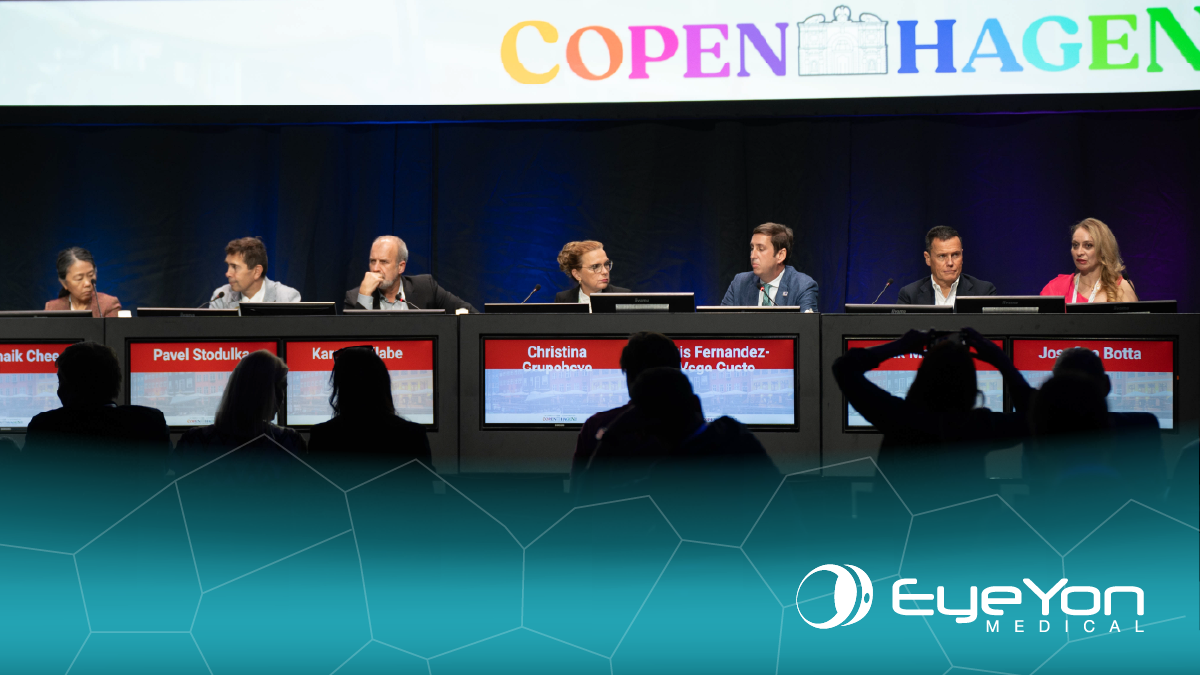
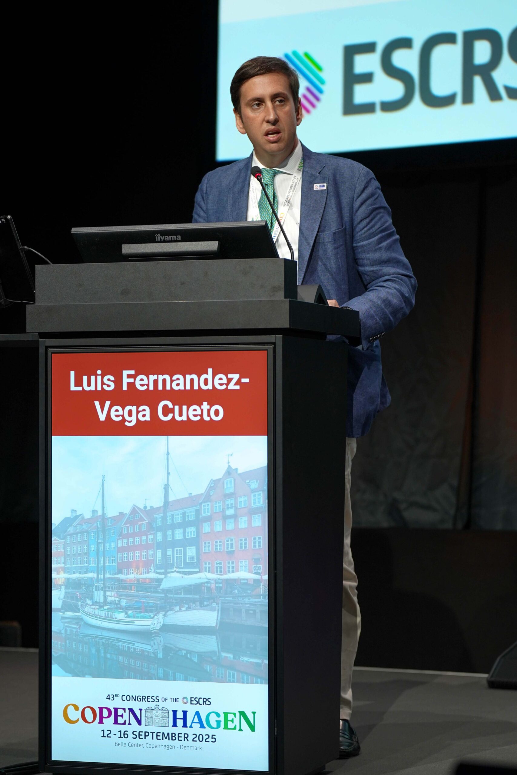
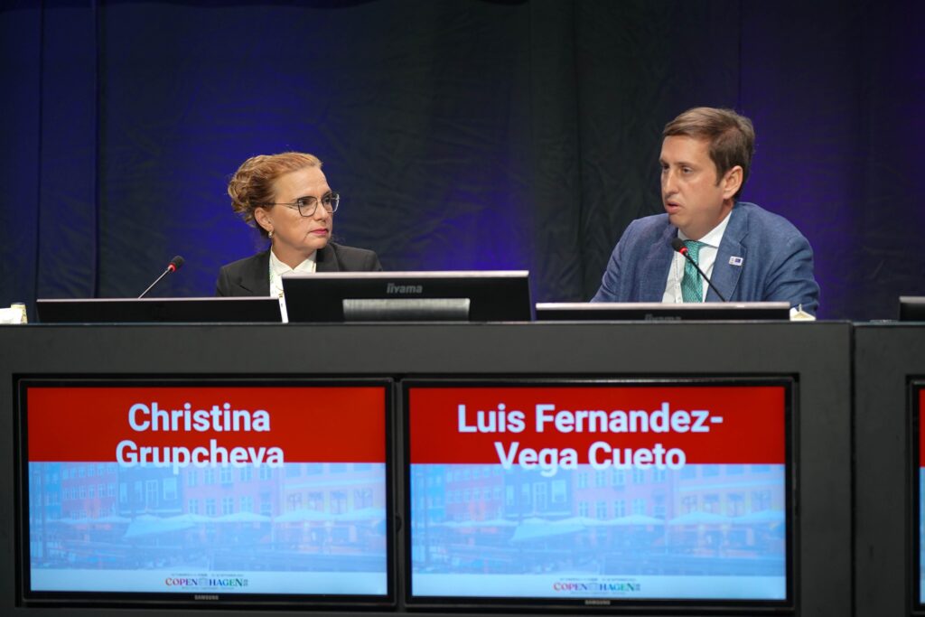
A highlight on Day 2 of the 43rd Congress of the European Society of Cataract and Refractive Surgeons (ESCRS 2025), is the EndoArt near-live surgery demonstration by Prof. Luis Fernández-Vega (Spain), during which he shared practical pearls such as synechiae management, endothelial removal under viscoelastic and optimal device positioning.
EndoArt was chosen as a primary treatment in such a case of a 74-year-old man who presented with bilateral pseudoexfoliative glaucoma with a functioning tube shunt and extensive anterior synechiae across the inferior corneal quadrant.
“We could have several surgical approaches, but we decided to do an EndoArt implant and release the inferior synechiae,” Prof. Fernández-Vega explained.
Such scenarios pose challenges for traditional endothelial keratoplasty, making EndoArt an attractive option due to its simplicity, availability, immediate corneal clarity and a significant reduction in central corneal thickness.
The EndoArt advantage
EndoArt is a biocompatible, optically clear hydrophilic acrylic implant marked with an ‘F’ for orientation. Measuring 6.5 mm in diameter, 6.8 mm in curvature and just 50 microns in thickness, “it creates a fluid-impermeable barrier to prevent the passage of aqueous humor into the corneal stroma, restoring homeostatic fluid balance,” Prof. Fernández-Vega explained.
Unlike Descemet membrane endothelial keratoplasty (DMEK) and Descemet stripping automated endothelial keratoplasty (DSAEK), which carry higher risks of rejection and failure, EndoArt enables immediate assessment of visual potential without waiting for donor tissue, a clear advantage in complex glaucoma cases.
Preparing the cornea
Prof. Fernández-Vega began the procedure by removing the anterior synechiae before implanting the EndoArt, carefully choosing a temporal approach to avoid interference with the superior tube and previous trabeculectomy.
“I’m going to remove the epithelium in order to have nicer visualization,” he said, as he began surgery. “We will do a paracentesis so we can manage the anterior chamber.”
Using viscoelastic alone, he cleared most of the adhesions in the inferior cornea, finishing the removal with a spatula to facilitate subsequent maneuvers. The main incision, 2.2 mm as in standard cataract surgery, was made temporally, though a superior approach could be used if needed.
Recognizing the complexity of the case, Prof. Fernández-Vega then removed the remaining endothelial layer under viscoelastic. “[In the past], we were doing it under air or with an anterior chamber maintainer,” he said. “Nowadays, we have very good results doing it under viscoelastic for complex cases like these.”
“It’s quite important to remove the remnants of endothelium from the recipient in order to help the EndoArt to have a good joining [adhere properly],” he added, carefully clearing fragments with a vitrectomy port to ensure smooth and secure implantation.
Implantation and positioning
Once the corneal bed was fully prepared, the EndoArt was inserted with ease, he positioned the device using a spatula, confirming correct orientation with intraoperative optical coherence tomography (OCT) and the “F” mark for alignment.
“We insert it with the spatula, but also do it with forceps. And now it’s opening circular,” he narrated. “And with these slow movements, we place the EndoArt right in the center of the cornea,” Prof. Fernández-Vega said. Once the device was positioned, he secured the main incision with sutures and adjusted the gas bubble to ensure complete adhesion.
Intraoperative OCT confirmed a 360-degree attachment, exactly as intended. “This is what we want to achieve in every EndoArt case, a device that sits perfectly still, with no areas of non-attachment,” he explained.
Twenty minutes later, repeat OCT imaging showed the implant was still firmly in place.
“The difference is striking,” Prof. Fernández-Vega noted. “The cornea is much clearer than at the beginning, and pachymetry confirms that it is also significantly thinner.”
Expert Q&A
During the Q&A session, Prof. Fernández-Vega addressed several key clinical questions.
In response to a question regarding whether he had experienced examples of detachment of the device, Prof. Fernández-Vega told the audience that at the beginning of their experience with EndoArt, if they had a detachment, they would go to the operating theatre to put in an air bubble every time, but now they only do that in cases where there is a significant detachment.
Regarding suture removal timing, he offered practical guidance based on experience with the novel device. “We think nowadays, of course, this was a novel device that we’ve been working with now for two years. And now the suggestion is to leave it at least six months. That would be our suggestion,” Prof. Fernández-Vega advised. “We had a case that I will present tomorrow where we removed the suture three months later, and we had a small detachment in that area. However, the center was clear. We left it like that, and the patient is fine.”
A new standard in endothelial care
EndoArt marks a new hope for patients who are not suitable for human tissue. “We have to think that it is not here to substitute human tissue, and that’s something that we have to have very clear, but it’s a new tool that extends all the indications,” Prof. Fernández-Vega concluded.
With rapid corneal clearing, secure attachment and stable outcomes, EndoArt empowers surgeons to restore vision even in the most challenging cases.
Editor’s Note: The 43rd Congress of the European Society of Cataract and Refractive Surgeons (ESCRS 2025) is being held from 12-16 September in Copenhagen, Denmark. Reporting for this story took place during the event. This content is intended exclusively for healthcare professionals. It is not intended for the general public. Products or therapies discussed may not be registered or approved in all jurisdictions, including Singapore.