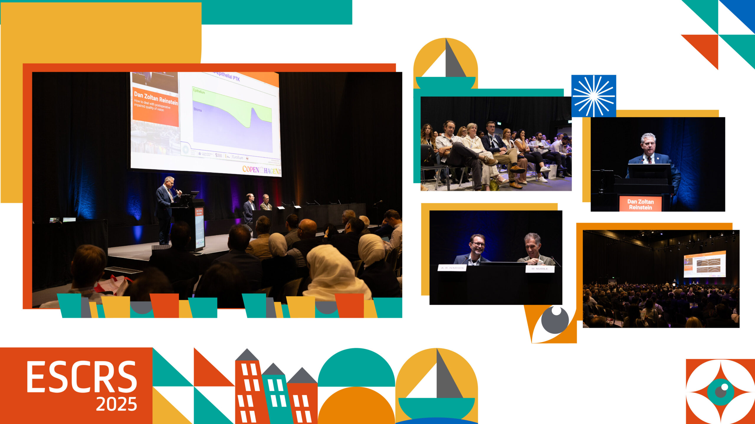
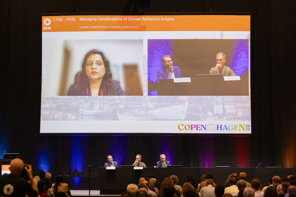
Corneal chaos decoded: strategies for navigating the trickiest refractive complications.
The opening day of the 43rd Congress of the European Society of Cataract and Refractive Surgeons (ESCRS 2025) featured a comprehensive Main Symposium where the “greatest hits” of refractive complications got their time in the spotlight.
Foggy flaps, wobbly suction, surprise black spots and even the dreaded ectasia. Each had its moment on stage, dissected with equal parts precision and pragmatism. With seasoned surgeons turning decades of scar tissue (figuratively, of course) into step-by-step wisdom, the audience got a front-row seat to what really matters: spotting trouble early, knowing your tools and remembering that sometimes the cornea writes its own plot twists.
READ MORE: Perfect Surgical Saves and Lessons from the Masters
Post-op complications
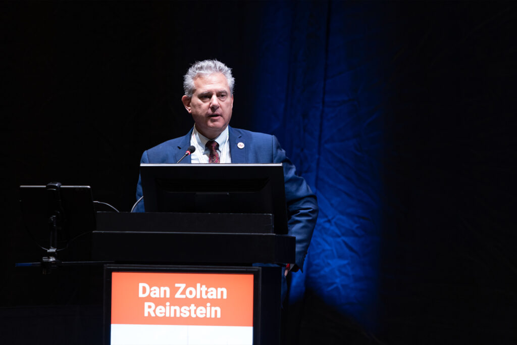
To say that diagnostics are important is an understatement. According to Prof. Dan Reinstein (United Kingdom), they’re the entire game when it comes to sorting out post-op quality-of-vision woes.
“The only reason people think I’m so good with refractive complications is that I started looking at the epithelium, stroma and flaps 30 years ago with high-frequency ultrasound,” he said. “I was taught by diagnostics, and the answer becomes very obvious as to what you have to do next.”
He unveiled his decision-making tree for troublesome corneas…essentially a “choose your own adventure” for irregularities. First up: ‘regularly irregular (global)’ corneas, where roughly 80% of the story is written in the topography and 20% in the epithelium. For these, topography-guided treatments do the job nicely.
But if you’re dealing with ‘irregularly irregular (local)’ corneas, it flips: 80% of the action is in the epithelium and just 20% in the topography, meaning trans-epithelial phototherapeutic keratectomy (TE-PTK) is your tool of choice.
READ MORE: How to Manage Complications in Phakic Lens Surgery—Lessons, Playbooks and Pearls
Femto-LASIK complications
Prof. Namrata Sharma (India) joined the session virtually (visa woes prevented her from being there in person) but that didn’t stop her from diving into the murky waters of femtosecond LASIK complications. The usual suspects on her list: opaque bubble layer (OBL), vertical gas breakthrough (VGB) and suction loss.
On OBL—the fog that clouds both surgeon vision and laser tracking—Prof. Sharma pointed out that its frequency can be anywhere between 5% and 72.6%, a range wide enough to make statisticians twitch. The silver lining? Newer-generation lasers are steadily cutting those odds down.
VGB, though far rarer (0.03% to 0.13%), calls for quick reflexes. “Either you stop immediately and release the foot pedal,” she advised, “or if it occurs prior to side cut and the flap is still intact, you repeat the laser from the opposite direction and give a deeper cut by 50 µm if the residual bed thickness allows.”
Then there’s suction loss, which is the LASIK equivalent of the rug being pulled out mid-procedure. Here, timing is everything. “If there is suction loss and the level of head treatment has been completed with no side cut, the procedure should be completed immediately,” Prof. Sharma noted. “But if it occurs during the side cut, do not lift the flap. Retreat the laser with a smaller flap, which is 0.5 mm smaller at a greater depth, which is 140 µm deeper.”
She wrapped things up with a reminder that the best way to manage complications is to stack the deck in your favor from the start: proper pre-op counseling, smooth docking technique and meticulous flap dissection and hydration.
KLEX-specific complications
Prof. Leonardo Mastropasqua (Italy) tackled complications unique to kerato-refractive lenticule extraction (KLEX), reminding the audience that while the procedure may look like LASIK’s younger cousin, its intraoperative hiccups are in a league of their own.
“KLEx has a unique spectrum of complications distinct from LASIK,” he noted. “Most are preventable and manageable with proper technique, surgical awareness and case selection.”
Many of these quirks stem from the usual troublemakers that come with lenticule dissection and extraction: suction loss, OBL, black spots, cap perforation, lenticule tears and the dreaded wrong-plane dissection.
READ MORE: Smart Tech in Refractive Surgery
Black spots, in particular, deserve extra caution. These tiny zones of complete photodisruption can cause outsized headaches if not handled properly.
According to Prof. Mastropasqua, the key is to determine their exact location and size within the stromal bed, since this helps the surgeon avoid dissecting directly through the spot. Instead, the dissection should be carried out around the affected area.
Looking ahead, he was optimistic about technology smoothing the path. “Emerging KLEx platforms promise to simplify dissection, improve technology [docking, centration, energy, stability, OCT imaging] and reduce complication rates.”
Corneal ectasia
Dr. Vance Thompson (United States) brought plenty of passion to his update on corneal ectasia, making it clear from the start: this isn’t just a LASIK issue.
He pointed to a 2021 review by Moshirfar and colleagues, which broke down ectasia rates as follows:1
- 20 per 100,000 eyes for photorefractive keratectomy (PRK)
- 90 per 100,000 for LASIK
- 11 per 100,000 for SMILE/KLEx
But numbers weren’t the only culprit in his crosshairs. Patient behavior got plenty of airtime, too. “You cannot get through a refractive surgery consultation…without me drilling you about, ‘Do you rub your eyes?’ or ‘Do you sleep on your tummy and push your eyes into a pillow?’” he stated emphatically. “I think it is one of the largest causes of post-refractive corneal surgery ectasia.”
For Dr. Thompson, the secret weapon against bad outcomes is epithelial mapping, paired with corneal topography. “There’s no way I’m ever going to do refractive surgery ever again without having epithelial mapping in combination with corneal topography,” he declared. “I consider the two married and extremely important,” pointing to data showing it altered surgical decisions in 16% of cases.2
Corneal haze in surface ablation
Dr. Sharita Siregar (Indonesia) took the not-so-clear issue of corneal haze after surface ablation, breaking down the “why” and the “what to do about it.”
“Haze can be created because of the alteration in corneal transparency caused by refractive surgery such as surface ablation,” she explained.
Several risk factors can tip the scales toward haze, including high ablation depth (myopia or hyperopia > 6 D, astigmatism > 3 D), stromal irregularities, severe dry eye, too much UV exposure and disruption of those all-important hemidesmosomes.
Dr. Siregar divided corneal haze into two categories: early onset (within three months) and late onset (beyond three months). The treatment playbook shifts accordingly. Early haze responds to prednisolone 1% every four hours, while late haze demands a stronger schedule of every two to three hours.
For the tougher cases, she didn’t mince words. “If we have deep haze, that means that we can do PTK or PRK using 0.02% MMC [mitomycin C],” Dr. Siregar advised. “As for superficial haze, we can do mechanical debridement and also use MMC combination.”
READ MORE: APACRS 2025 Tackles Tough Cases in Cataract and Refractive Surgery
Corneal surgery for refractive complications
Prof. Dr. Jose Guell (Spain) had the honor of closing the symposium with a topic nobody wants to talk about, but everyone eventually has to: corneal surgery for severe refractive complications.
While he reassured the audience that such cases are rare—fewer than 0.03% of patients end up needing keratoplasty after refractive surgery—the sheer number of procedures worldwide means tricky cases inevitably land on a surgeon’s desk.
Prof. Guell divided the complications into two camps: anterior and posterior corneal problems. For the anterior group, he stressed a crucial step before reaching for the scalpel: always check best-corrected vision with rigid gas permeable lenses first. “In most cases, we might deal with them with rigid contact lenses,” he said, showing cases of irregular astigmatism that scleral lenses had tamed beautifully.
When surgery does become unavoidable, Prof. Guell explained that his preferred strategy for flap repair is to remove all of the epithelium and then elevate the flap. In these cases, there is no need to stretch the flap; simply removing the epithelium is sufficient.
For the truly tough cases, he turned to the heavy artillery: deep anterior lamellar keratoplasty (DALK) and penetrating keratoplasty (PKP). “In some cases, like high irregularity post radial glia without the capability of using contact lenses, we need to do full thickness,” he explained.
On the posterior side, Prof. Guell noted that while only around 10% of phakic IOL patients may need transplantation within 15 years (with 4% to 5% requiring endothelial transplants), the rising popularity of these implants means the absolute number of such cases is set to grow.
READ MORE: The Corneal Comeback
The takeaway
At the end of the session, one thing was clear: refractive complications may be rare, but they never fail to keep surgeons on their toes. The take home message? Arm yourself with diagnostics, know your corneal quirks and keep your toolbox ready.
Dive into all the stories from ESCRS 2025 with our daily coverage.

Editor’s Note: The 43rd Congress of the European Society of Cataract and Refractive Surgeons (ESCRS 2025) is being held from 12-16 September in Copenhagen, Denmark. Reporting for this story took place during the event. This content is intended exclusively for healthcare professionals. It is not intended for the general public. Products or therapies discussed may not be registered or approved in all jurisdictions, including Singapore.
References
- Moshirfar M, Tukan AN, Bundogji N, et al. Ectasia after corneal refractive surgery: A systematic review. Ophthalmol Ther. 2021;10(4):753-776.
- Asroui L, Dupps WJ Jr, Randleman JB. Determining the utility of epithelial thickness mapping in refractive surgery evaluations. Am J Ophthalmol. 2022;240:125-134.