Top surgeons from Europe joined APAC regional experts to share must-have techniques for managing posterior capsule tears in an APAO-AIOC 2025 Day 3 symposium.
Posterior capsule tears (PCTs) are one of the most dreaded complications in cataract surgery. But lucky for us, some of Europe and Asia’s most preeminent surgeons have our backs.
At the 40th Asia-Pacific Academy of Ophthalmology Congress, held in conjunction with the 83rd Annual Conference of the All India Ophthalmological Society (APAO-AIOC 2025), the European Society of Cataract and Refractive Surgeons (ESCRS) co-hosted a session packed with contemporary surgical takes on posterior capsule tear management.
This collaborative session brought together global cataract experts from Europe and Asia to share their approaches to this significant complication. The panel featured Prof. Marie José Tassignon (Belgium), developer of the bag-in-the-lens technique; Dr. Rohit Om Parkash (India); ESCRS President-Elect Prof. Burkhard Dick (Germany); Dr. Vladimir Pfeifer (Slovenia); Prof. Thomas Kohnen (Germany), Chair of Ophthalmology at Goethe University Frankfurt; and Dr. Amar Agarwal (India), Chairman of Dr. Agarwal’s Eye Hospital.
Understanding the anatomy
Prof. Tassignon (Belgium) focused on the importance of understanding posterior segment anatomy, and especially the ligament of Wieger and anterior vitreous detachment (AVD), as critical for recognizing the potential for capsular tears.
“[Understanding] more about the mechanics [of the eye] behind the posterior capsule is important,” she said, “and this is really not a black box. It should be something that everybody should know about.”
Prof. Tassignon’s key point for using anatomical knowledge to up your PCT game? Getting skilled at recognizing signs of AVD during surgery—such as lens fragments in intact vitreous—can alert surgeons to a potentially fragile posterior capsule, and help them take necessary actions sooner.
Parkash’s PCT pearls
Dr. Parkash’ presentation was chock full of practical tips for all surgeons looking to manage PCTs in their operating room.
He started by pointing out a critical distinction. “We have to realize that the anterior vitreous face and posterior capsule structure, as Marie has just shown, are 2 different things,” he explained.
One of his top revelations was that high fluid parameters and turbulence should be avoided at all costs, as they can lead to complications even with an intact capsule.
To this end, Dr. Parkash outlined several strategies for implementing anterior hyaloid face-friendly practices, including proper preoperative scanning for preexisting posterior capsule defects and calibrated incisions for stable chamber settings.
Taking a measured approach to phacoemulsification is another key strategy for avoiding PCTs, according to Dr. Parkash. “You should have active fluidics… and slow motion phacoemulsification, because that creates a situation wherein there is a minimal pressure difference between the anterior and posterior segments,” he said.
Through case demonstrations, he showed how to manage challenging scenarios including fibrotic cataracts and posterior polar cataracts with preexisting defects. His approach includes using low infusion pressure (IOP at 20), avoiding chamber fluctuations, and utilizing nuclear fragments or IOLs as scaffolds. “With a low infusion at 20 IOP, the posterior capsule is not being pushed behind. The chances of the anterior hyaloid face being ruptured is small,” he said in one surgical analysis.
In a particularly instructive case of posterior polar cataract with a preexisting defect, he demonstrated how proper chamber settings prevent nuclear fragments from descending posteriorly: “Even with a posterior capsule rupture, [nuclear fragments] won’t descend. This shows that with the anterior hyaloid face intact, you can create a situation where the anterior hyaloid face doesn’t get broken whatsoever.”
Limbal vs. pars plana vitrectomy
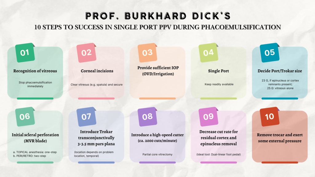
Prof. Dick (Germany) discussed a choice every surgeon faces when performing a vitrectomy during PCTs: going with a pars plana vitrectomy (PPV) or a limbal (LAV) approach.
“The posterior approach is better than anterior,” he said, pointing to lower incidence of post-operative retinal detachment, cystic macular edema, IOL instability and endophthalmitis.
His presentation included a detailed, 10-step protocol for a single port pars plana vitrectomy during phacoemulsification.
PCT prevention and immediate management
Dr. Pfeifer’s (Slovenia) presentation was themed around how to prevent PCTs in the first place, and the importance of swift action once they do occur.
On the prevention front, Dr. Pfeifer believes that capsulorhexis is “the most important phase of phaco” for preventing tears. His preferred technique keeps the phaco tip centered while using a second instrument to bring nuclear material forward, reducing posterior capsule stress.
When a tear occurs, Dr. Pfeifer urges immediate action—much like Prof. Dick.
“Stop perfusion,” he said. “Lead the phaco tip in and inject the viscoelastic or whatever is at hand. Then you have a stable situation, and you can think.”
IOL exchange considerations
As European Editor at the Journal of Cataract and Refractive Surgery, Prof. Kohnen echoed Dr. Pfeifer’s sentiments on the importance of the capsulorhexis by providing data. According to his dissertation, in the two years (1986-1988) since the change to capsulorhexis from the can-opener technique, PCT rates decreased from 8.4% to 4.8%.*
Like Dr. Pfeifer, Prof. Kohnen also believes that prevention is the best medicine: maintaining the anterior capsulorhexis in modern cataract is as crucial as ever.
As for Prof. Kohnen’s top tip, it’s all about cutting his lens into pieces—but not as his last resort.
He demonstrated multiple explantation techniques for cutting IOLs, all while emphasizing the importance of minimizing incision size. But in the end, despite advancements like the capsulorhexis, the most important thing is vigilance. “Be prepared for a posterior capsule [tear] at all times,” he said.
IOL scaffold technique
To conclude the session, Dr. Agarwal (India) demonstrated an important technique for PCTs: IOL scaffolding. “You are creating your own posterior capsule to emulsify the nucleus and put the lens into the sulcus—if you have a good rhexis,” he said.
This approach—demonsrated by placing a three-piece IOL with haptics above the iris—creates a barrier, preventing nuclear fragments from descending posteriorly.
Practical takeaways
So what were the key takeaways from the symposium? We sum it all up here.
- Understand posterior segment anatomy and recognize signs of anterior vitreous detachment
- Perfect capsulorhexis technique for maximum prevention
- Consider immediate stabilization with viscoelastic when a tear occurs
- Pars plana vitrectomy could be the key to improved PCT outcomes
- The IOL scaffold technique could help prevent nuclear fragments from falling posteriorly
*Friedrich-Wilhelms Universität Bonn, Dissertation: „Kapsel- und Zonularupturen als Komplikationen der Kataraktoperation mit Phakoemulsifikation“ 06.06.1989, Gesamturteil: sehr gut
Editor’s Note: Reporting for this story took place during the 40th Congress of the Asia-Pacific Academy of Ophthalmology (APAO 2025), being held in conjunction with the 83rd Annual Conference of the All India Ophthalmological Society (AIOC 2025) from 3-6 April in New Delhi, India.