When cataracts collide with complex eyes, strategy is survival. This ASCRS 2025 symposium served the ultimate battle plan for tricky surgeries.
Cataract surgery gets a lot trickier when other eye diseases join the party—and that’s exactly what the Cataract Crossover Symposium tackled on Day 3 at the American Society of Cataract and Refractive Surgery Annual Meeting (ASCRS 2025).
Moderated by a powerhouse trio—Drs. Cathleen M. McCabe, Marjan Farid and Zaina Al-Mohtaseb—the session explored how to navigate cataracts when the patient brings along lid abnormalities, retinal disease, glaucoma, neuro-ophthalmic challenges or corneal disorders.
From pre-op planning to post-op curveballs, the session was packed with practical pearls for tackling complex eyes.
Periocular pitfalls of cataract surgery
Prof. Dr. Seanna Grob (United States) turned the spotlight on an often-overlooked player: the eyelids. As she reminded the crowd, “The eyelids in orbit are really the protectors of the globe. If you don’t have the eyelids, the cornea will just kind of melt.”
From ptosis to facial nerve palsy and thyroid eye disease, she showed how periocular anatomy can quietly sabotage what looks like a straightforward cataract case. Even something seemingly minor—like ptosis—can skew your pre-op measurements: “If ptosis is significant, it may actually affect your preoperative calculations.”
And post-op? Surgeons should stay alert. Ptosis after surgery isn’t rare—rates can range from under 5% to as high as 30%. Bottom line: success isn’t just about the lens—it’s about respecting the whole ocular surface system.
WATCH NOW: Simplifying Complex Cataract Cases with the Right Techniques
Patients with neuro-ophthalmic disease
Dr. Vivek R. Patel (United States) then brought the room to full attention at ASCRS 2025 as he unpacked one of cataract surgery’s trickiest terrains: patients with neuro-ophthalmic disease.
With his engaging style, Dr. Patel walked the audience through case after fascinating case, showing that not all vision loss is about the cataract—and why surgeons need to dig deeper.
First up: patients with optic nerve damage from multiple sclerosis or past optic neuritis. Even if the cataract is obvious, “OCT of the optic nerve and ganglion cell analysis can be super helpful,” he advised. Thinning of the ganglion cell layer, particularly below 75 microns, can hint at permanent visual field loss—even if the lens looks like the main culprit.
Then came a show-stopping reminder: visual fields still matter. In one case, a seemingly ordinary cataract patient actually had a massive prolactinoma, caught only thanks to a suspicious visual field test and subtle ganglion cell layer changes.
Dr. Patel also tackled the much-debated question about cataract surgery increasing the risk of ischemic optic neuropathy (ION). His take? Modern surgical techniques have significantly lowered that risk, but a careful conversation with patients—especially those with “disk-at-risk” anatomy—is still essential.
Glaucoma patients deserve more
Dr. Zarmeena Vendal (United States) didn’t hold back when diving into the complexities of cataract surgery in glaucoma patients at ASCRS 2025. She encouraged surgeons to rethink outdated limits when treating glaucoma patients needing cataract surgery.
READ MORE: Cataracts vs Glaucoma: What You Need to Know
She walked through her approach to lens selection: EDOF lenses like Alcon’s Vivity and J&J’s TECNIS Symfony are strong choices for mild to moderate glaucoma cases, providing crisp distance and functional intermediate vision without sacrificing contrast sensitivity. She’s also a big fan of Light Adjustable Lenses (LAL) for patients with tricky biometry.
READ MORE: RxSight Brings the Party and Precision to ASCRS 2025 with LAL
But for Dr. Vendal, mastering MIGS is no longer optional. “If we’re going to dabble in the world of advanced technology lenses, it behooves us to absolutely also be comfortable doing MIGS at the same time.”
With smart strategy, she argued, we can give glaucoma patients freedom from glasses and better control over their disease—all at once.
Watch out—retinal trouble can hide
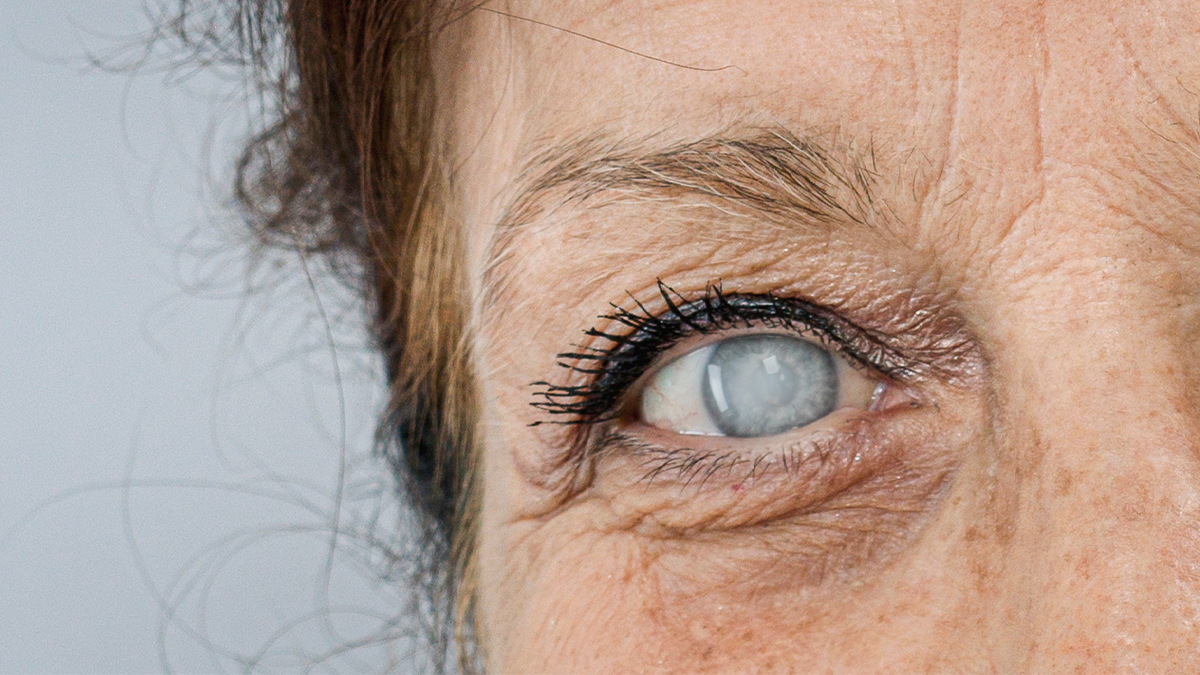
When it comes to cataract surgery in patients with retinal disease, Dr. Mitul Mehta (United States) didn’t mince words: start with a pre-op OCT—or prepare for trouble. “Sometimes the cataract is pretty bad and you can’t see subtle pathology,” he said, urging surgeons not to skip that crucial imaging step.
Dr. Mehta walked through cases where multifocal lenses were mistakenly implanted in eyes with hidden retinal problems like macular traction and epiretinal membranes—mistakes that could have been caught early with an OCT. He made it clear: “Multifocal lenses are a no-go zone for retinal pathology.”
For patients with stable mild disease, he advocated for careful choices like EDOF lenses or LALs, depending on the individual eye. And when it comes to diabetic retinopathy, even without macular edema, pre-op anti-VEGF injections can be smart insurance.
His advice? Show patients their OCTs, manage expectations upfront and don’t assume the retinal status just by slit lamp exam. “If you don’t look, you’re not going to know,” he said, reminding the audience that retinal diseases can sneak up silently—and post-op surprises are no fun for anyone.
Smooth sailing through corneal disease
Closing the symposium, Dr. Zeba A. Syed (United States) tackled cataract surgery when the cornea isn’t playing nice. “Corneal diseases do affect IOL selection. Cataract surgery has the potential to contribute to progression of preexisting corneal diseases… and of course, it limits visual outcomes and affects our counseling.”
Dr. Syed took the audience on a fast-paced tour through common corneal challenges, starting with dry eye syndrome, which she called a major culprit in bad biometry.
“We know that it affects the topography. It prevents precise keratometry readings for accurate IOL calculation,” she explained, emphasizing the risks when planning for premium lenses.
READ MORE: Pentacam Cornea OCT Offers Synergy for the Future of Corneal Assessment
She also shared pearls on anterior basement membrane dystrophy (ABMD), stressing the importance of treating irregular astigmatism early—and waiting three months post-treatment before biometry.
For HSV keratitis, Dr. Syed recommends a three-month inactivity period before surgery and starting prophylactic antivirals. In keratoconus, she flagged the importance of stability checks and cautious use of toric lenses.
When tackling Fuchs’ dystrophy, she often pairs cataract surgery with DMEK—and always plans for a hyperopic shift after endothelial transplants.
The future of cataract surgery is crossover
The Cataract Crossover Symposium wasn’t just another stop on the ASCRS 2025 schedule—it was a rallying cry for surgeons ready to raise the bar. Whether it’s lids, nerves, retinas or corneas, the old rules no longer apply. Today’s cataract surgery demands fearless planning, smarter tools and a new level of clinical swagger. If you’re not crossing specialties, you’re falling behind. The future is complex—and it’s already here.
READ NOW: Get the latest news from ASCRS 2025 with our daily updates here!
Editor’s Note: Reporting for this story took place during the annual meeting of the American Society of Cataract and Refractive Surgery (ASCRS 2025) being held from 25-28 April in Los Angeles, California, United States.